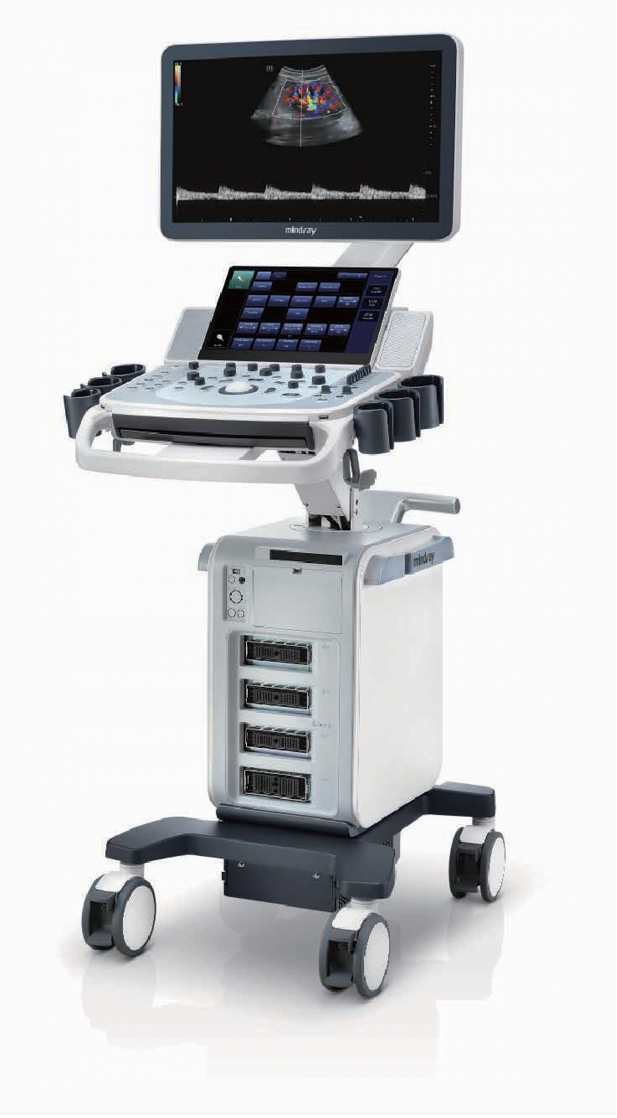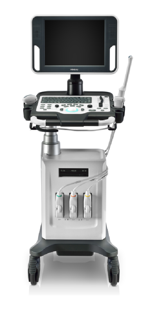


Current low-end ultrasound systems typically allow only basic diagnosis but lack advanced functionalities. Now, with competitive pricing in the ultrasound market, the DC-30 machine is a perfect solution for high-quality imaging performance, featuring additional functions like Auto IMT, iScape, Natural Touch Elastography, UWN Contrast Imaging, and Doppler Tissue Imaging.
PRODUCT FEATURES
Performance
PSHTM (Phase Shift Harmonic Imaging)
Phase Shift Harmonic Imaging technology enhances contrast resolution, providing ultrasound images with high resolution and reduced image noise.
iBeamTM
Enables the use of multiple ultrasound scan angles to create a single image, enhancing contrast resolution and image display.
iClearTM
Improves image quality based on automatic tissue structure detection:
Clear and seamless edges
Smooth and uniform tissues
Empty echo areas free from artifacts
Multiple Beam Formation
Increases transmitted ultrasound beams by four times, leading to excellent temporal resolution and higher frame rates.
Natural Touch Elastography
Based on Mindray's latest patented technology, Natural Touch Elastography quantitatively analyzes tissue elasticity with a single surface, providing high-sensitivity stiffness assessment in applications like thyroid, breast, and gynecology.
iScapeTM
Expands the image to adjacent anatomical structures, providing an overview of the structure to be examined.
ExFOV
Enhances diagnostic capabilities through Extra Point Of View mode for anatomical structure expansion on all convex and linear probes.
B-SteerTM
A feature for deep tissue biopsy: helps adjust the scan line for better visualization of needles, nerves, and small blood vessels.
Free XrosTM
Obtain accurate anatomical observations by placing sample lines at any angle.
TDI
Doppler tissue imaging enables quantitative assessment of local motion and cardiac function, providing complete TDI modes for direct and faster diagnoses.
Workflow
iScanHelper
Integrated specialized guidance software:
Illustrates anatomical diagrams including schematic images and ultrasound
Compares standard ultrasound images with real-time scans
Guides image slices to provide appropriate patient positions and probe placements
Tips on scanning skills and diagnostic information
Auto IMT
Automatically measures the thickness of arteries before and after providing accurate carotid artery conditions.